
Aesthetic clinical crown augmentation with Ceramic Drill: case report
Learn about the ceramic drill clinical crown augmentation technique, an effective alternative for treating gummy smiles with immediate cauterization and excellent aesthetic stability. Check out the summary of this award-winning case report from the 42nd CIOSP.

Authors: Caio Vinicius G. Roman-Torres, Alvaro Luiz M. P. Barbora,
Luana Campos, Humberto O. S. Filho, William C. Brandt.
Reference: Paper presented at the 42nd CIOSP, winner of 1st place in the Clinical Forum.
The search for facial harmony inevitably involves a balanced smile. In modern dentistry, aesthetics is not just about the color or shape of the teeth, but about the proportional relationship between the lips, gums and teeth. When this harmony is disrupted by excessive exposure of gum tissue – the so-called “gummy smile” – the impact on the patient’s self-esteem can be significant.
In this article, we detail an award-winning case report from the 42nd CIOSP that explores the use of ceramic drills as an alternative to traditional scalpels in clinical crown augmentation.
The clinical case
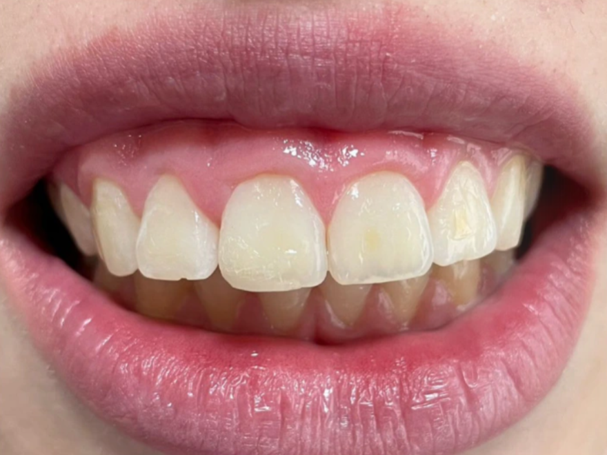
A smile that is considered aesthetically acceptable generally shows all the crowns of the upper incisors and around 1 mm of gingiva. Exposures of more than 3 mm are classified as a high or gummy smile.
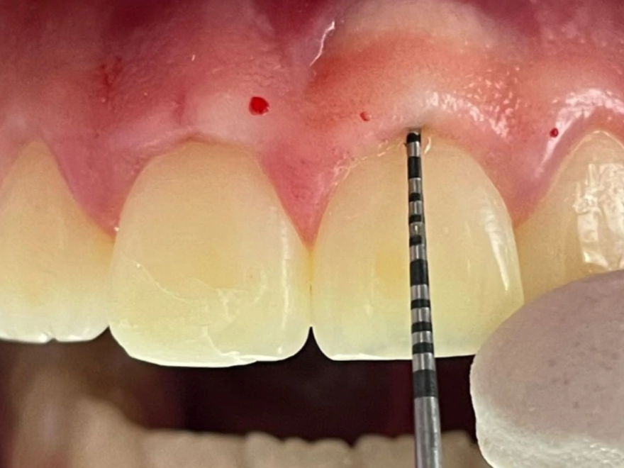
One of the common causes of this insufficient crown length is Altered Passive Eruption (APE). This is a condition where the gingival margins do not recede to the level of the cemento-enamel junction during tooth eruption. To correct this condition, clinical crown augmentation emerges as the ideal surgical procedure to expose the necessary tooth structure.
Technique used: the Ceramic Drill
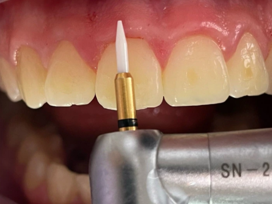
The great innovation of this clinical protocol lies in the replacement of conventional cutting instruments with the Soft Bur ceramic rotary bur (Microdont®). Although scalpel blades are the gold standard due to their cost and precision, they require meticulous manipulation and often result in bleeding that can hinder refined visualization of the operative field. The use of the ceramic drill changes this dynamic through three fundamental pillars:
- Rotating system: Use of 400 rpm without serum irrigation.
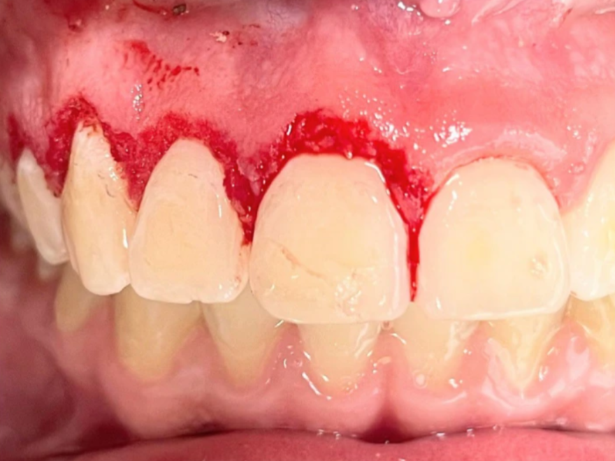
- Immediate Cauterization: The heat produced by the friction of the drill results in tissue coagulation and minimal bleeding.
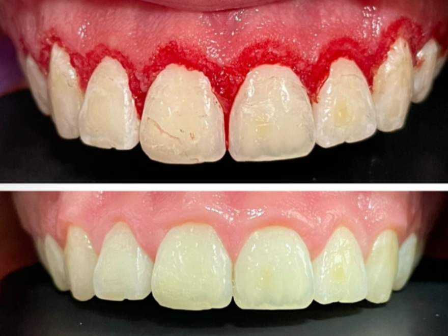
- Single Time: The design of the drill allows gingivectomy (height removal) and gingivoplasty (width adjustment) to be performed simultaneously.
Results and clinical conclusion
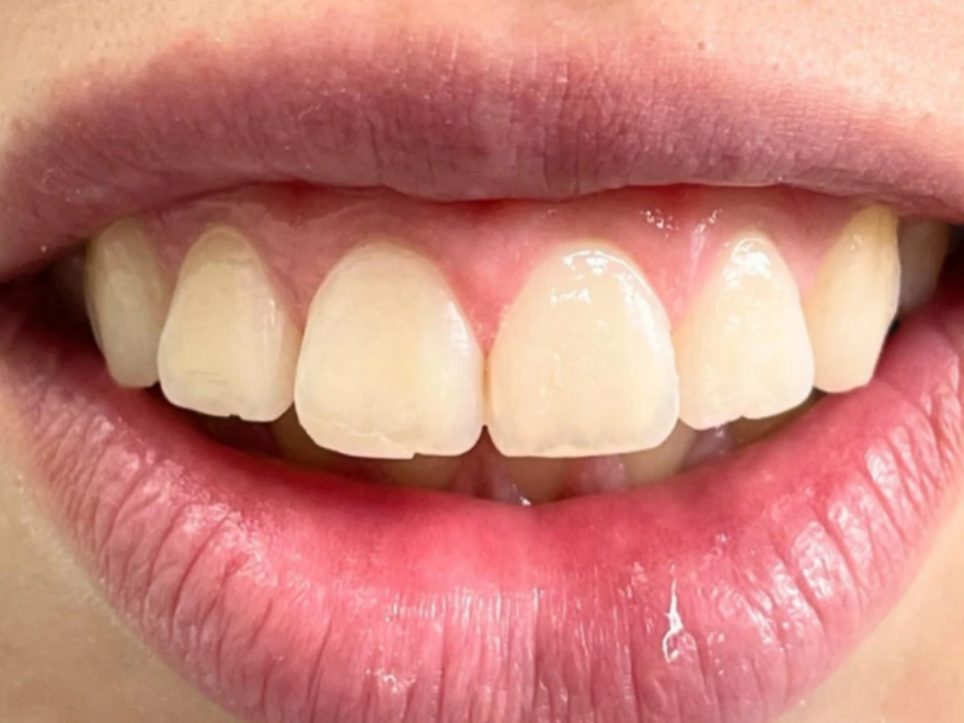
The 180-day follow-up confirmed the effectiveness of the technique. At all returns (2, 5, 7, 30, 90 and 180 days), the patient showed periodontal health and total stability of the gingival margins.
Why consider the ceramic drill?
Although scalpel blades continue to be the gold standard, ceramic drills have emerged as a lower-cost and easier-to-use alternative, since they take advantage of the dentist’s familiarity with high-precision rotary instruments, offering several competitive advantages:
- Cost-effective: Lower than lasers and electrocautery.
- Learning curve: Easy to use for dentists already used to rotary instruments.
- Efficiency: Allows refinement of aesthetic details with greater control in healthy tissue.
This case demonstrates that the ceramic drill is a viable and efficient tool for clinicians seeking stable aesthetic results and a cleaner trans-operative procedure through cauterization.
Authors: Caio Vinicius G. Roman-Torres, et al.